Spots on children’s skin
Spots on children’s skin

This post was written by Dr. Pleimes, a specialist in pediatrics, dermatology and allergology
Click here to read further information about Dr. Pleimes.
Spots on children’s skin can often cause concern for parents. From small freckles to light brown café-au-lait spots and striking port-wine stains – the range of skin changes is vast and can sometimes raise questions. Most of these spots are harmless, but it’s important to keep an eye on certain changes and seek medical advice if necessary. In this article, you’ll learn about the types of spots that can appear on children’s skin, how they look, and when a medical check-up is advisable.
What do spots on children’s skin look like?
Even in children, different types of spots can appear on the skin. Some are present from birth, while others develop during childhood.
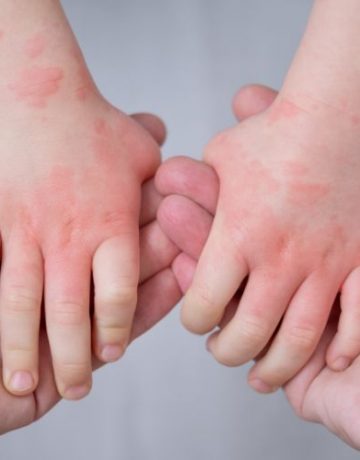
Spots may contain more or less pigment than the surrounding healthy skin. This can result in white (less pigment) or brown (more pigment) spots. If the pigment is located in deeper skin layers, the colour may appear more greyish-blue. The skin can also have a different colour if an area of skin is supplied with more or fewer small blood vessels than usual. Spots with less blood flow appear lighter, while those with increased blood flow may be red.
Spots caused by enlarged blood capillaries
Stork bite
A common harmless spot on the neck of children is the so-called stork bite. This spot, usually present from birth, is caused by a slight, very superficial enlargement and multiplication of capillaries, the smallest blood vessels. The stork bite often remains on the neck for life, while those on the eyelids or forehead usually fade during childhood.
Port-wine stain
Port-wine stains are also flat, reddish to violet spots that are initially at skin level. They, too, consist of enlarged blood capillaries and are present from birth. Port-wine stains tend to be more pronounced than stork bites and often appear on one side of the body, unlike stork bites, which usually show symmetrically along the midline. Port-wine stains vary in size, often measuring several centimetres. They persist without treatment and may thicken or darken over time.

Spots caused by skin pigmentation
Freckles
Light brown, non-raised spots can appear as small pigmentations in the form of freckles. These usually occur in individuals with fair, sun-sensitive skin types.
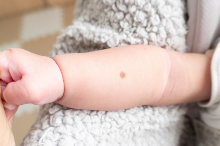
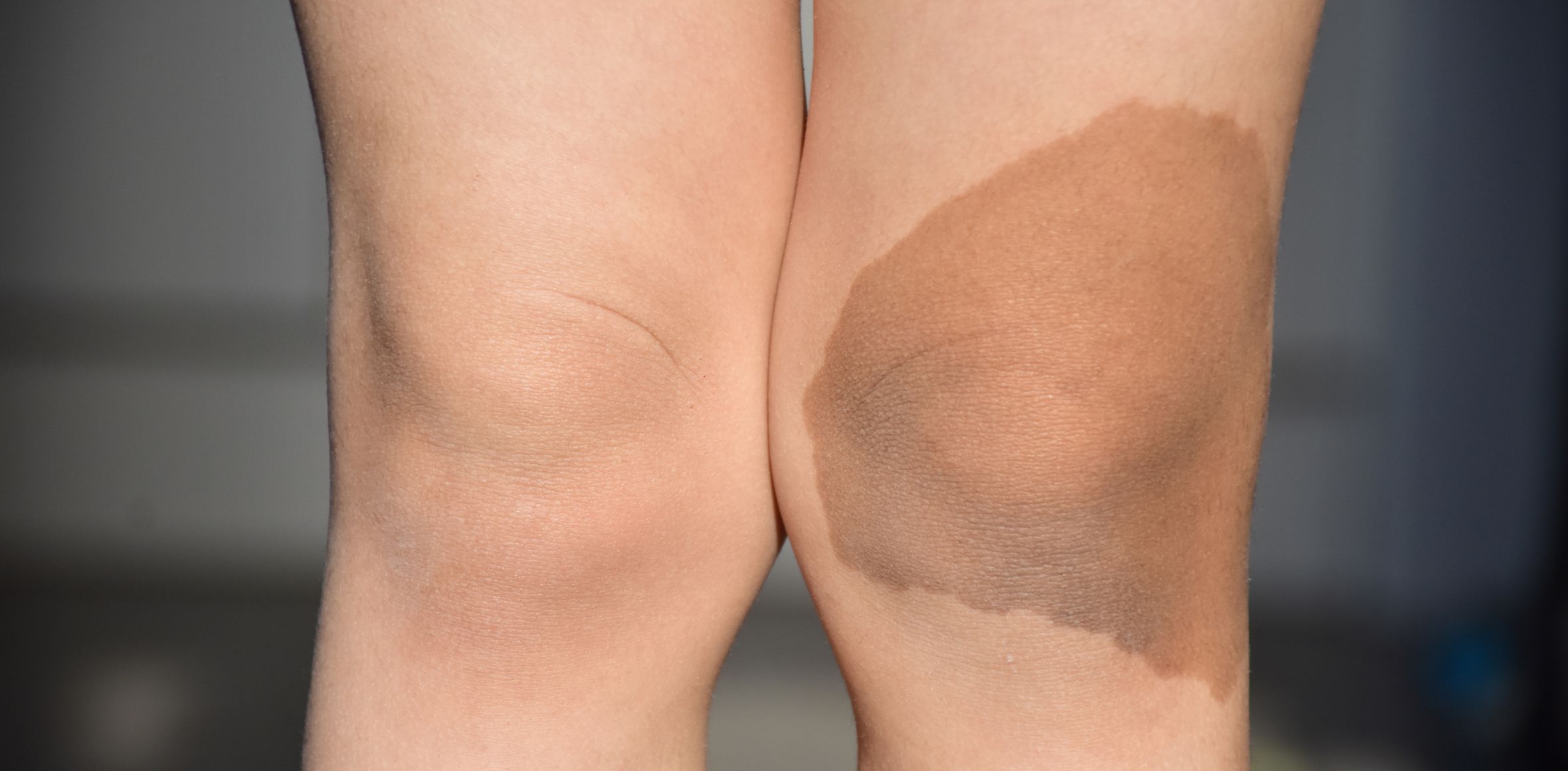
Café-au-lait spots
Large, often several centimetres in size, light brown spots sometimes appear as so-called café-au-lait spots. They are harmless when occurring singly. However, if a child has multiple or very large pigmented spots, they should be examined by a paediatrician.
Naevus depigmentosus
White spots that appear in young children during their first years are often already present at birth but only become noticeable as the surrounding skin becomes more pigmented. A harmless example of this is a naevus depigmentosus, a less pigmented spot that persists for life and grows proportionally with the body.
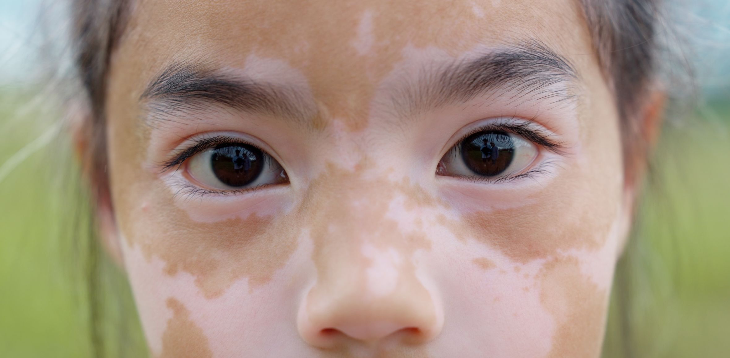
Vitiligo (white spot disease)
If new white spots keep appearing and light spots slowly change shape, this may suggest an acquired pigmentation issue. This could be vitiligo, a condition where the body mildly and harmlessly reacts against its own pigment cells, causing them to stop functioning properly. This results in the formation of light, depigmented spots. Vitiligo is generally harmless, but school-aged children may be affected by the visible lesions. While the condition cannot be cured at its root, treatment options are available for bothersome spots, and further examinations may be needed.
The difference between birthmarks and moles in children
Leave a comment
Changes in children’s skin
Skin rashes in children are quite common and can have various causes. The causes range from infections with viruses, bacteria, fungi and parasites to infections with viruses that cause warts. In this article, you can find out how the various skin changes manifest themselves and how they progress.
Find out more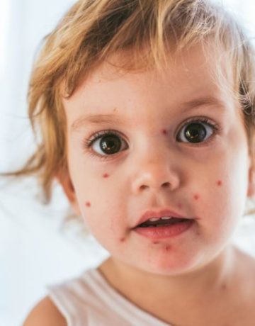
Viral exanthema in childhood
The term exanthema refers to a skin rash that typically affects larger areas of the body. In this article, you will find out what causes exanthema in children, what effects it has and how to differentiate between the various exanthema diseases.
Find out more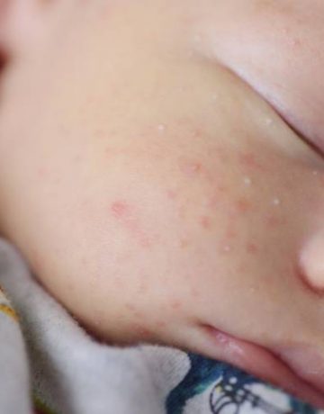
Neonatal acne: when pimples appear on baby’s skin
Shortly after birth, newborns may develop a variety of skin reactions, most of which are harmless. Three common skin reactions are: newborn acne, pityrosporum folliculitis and seborrheic dermatitis. In this article you will learn how these skin reactions differ from each other and how they can be treated best.
Find out more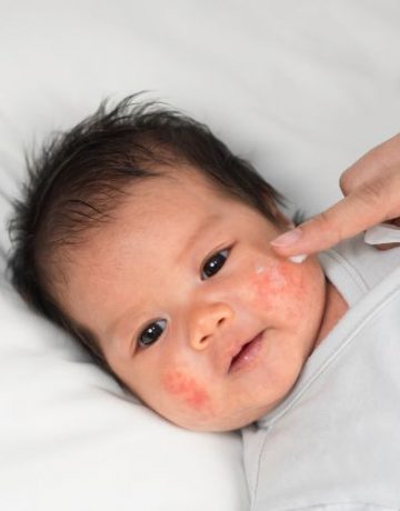
Atopic dermatitis in infants
In this article you will learn what neurodermatitis / atopic dermatitis or atopic eczema means, which causes are responsible and which skin areas are particularly affected. In addition, you will receive a detailed description and some tips on how atopic dermatitis can be treated in infants.
Find out more
Baby skin, detergents and textiles
In this article you will learn which detergent should be used to wash laundry and baby clothes so as not to damage the sensitive baby skin, whether it makes sense to use fabric softener and which textiles are suitable for baby clothes.
Find out more
Cleaning and caring for baby skin – from head to toe
In this post, you’ll learn how to properly clean (e.g. correct navel care), nurse and diaper your baby.
Find out more
Baby skin in the first weeks of life
In the first months of life, newborns may experience increased skin changes, such as neonatal exanthema or neonatal jaundice. In this article, you will learn how these skin changes are treated and whether they can be dangerous for your baby.
Find out more
Comments (0)